Spaces with a purpose
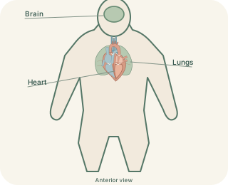
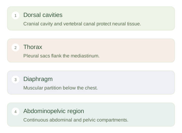
The body contains internal spaces that protect organs and allow them to move. The dorsal group includes the cranial cavity inside the skull and the vertebral canal within the spine. Together these protect the brain and spinal cord. The ventral group includes the thoracic and abdominopelvic cavities. These are useful organising regions rather than empty containers: organs, vessels, connective tissues and small amounts of fluid occupy them. Not every organ lies inside a fluid-lined space, and the membranes around different structures have different names.
Body Cavities and Organ Relationships
 Open diagram larger ↗
Chest, abdomen and pelvis
The diaphragm separates the thorax from the abdomen. In the chest, each lung has its own pleural sac. Between the lungs is the mediastinum, containing the heart, major vessels, oesophagus and other structures. The heart is enclosed by the pericardium. Below the diaphragm, the abdominal cavity contains much of the digestive system. The pelvic cavity continues inferiorly without a separating wall and contains structures including the bladder and rectum. Some organs, such as the kidneys, lie behind the main abdominal lining and are called .
Layers that slide
A serous membrane has a layer associated with the body wall and a layer covering an organ. Between these layers is a very thin, fluid-containing potential space. The fluid helps surfaces glide, for example as lungs expand or the heart beats. The pleural cavity is therefore the narrow space between pleural layers, not another name for the lung itself. Knowing these relationships helps explain why fluid can collect around an organ without collecting inside it. A regional map should distinguish the organ, its covering and the surrounding compartment.