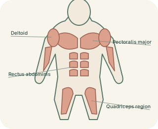
Skeletal muscle moves and supports
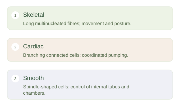
usually attaches to bones through tendons, although some muscles attach to skin or other tissues. Its long cells contain many nuclei and show stripes, or striations, because contractile proteins are arranged in repeating units. Motor neurons stimulate these fibres. We can voluntarily control many skeletal movements, but postural adjustments and breathing also involve automatic control. Skeletal muscle produces movement, helps stabilise joints and generates heat. Calling it voluntary is therefore a useful shorthand, not a claim that every contraction follows a conscious decision.
Skeletal, Cardiac and Smooth Muscle
 Open diagram larger ↗
Cardiac muscle pumps blood
forms the working wall of the heart. Its branching cells are striated and linked at specialised junctions called intercalated discs. Mechanical connections hold cells together, while gap junctions help electrical activity spread. Specialised cardiac cells can generate rhythmic activity, which the autonomic nervous system and hormones modify. The heart does not need a separate conscious instruction for each beat. Its organisation supports coordinated contractions that move blood through a continuous circuit. Unlike , cardiac muscle normally has very limited capacity to replace a large loss of functional cells.
Smooth muscle adjusts hollow organs
occurs in structures including blood vessels, airways, the gut and bladder. Its cells lack visible striations because their contractile proteins have a different arrangement. They can maintain force efficiently and often respond to stretch, local chemicals, hormones and autonomic input. Narrowing a small artery changes resistance to flow, while coordinated gut contractions move contents along. Smooth muscle is not simply a weaker version of : it is adapted to sustained control of internal tubes and chambers. Comparing these tissues shows how different cellular arrangements solve distinct problems involving force, timing and energy use.