Two pumps in series
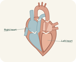
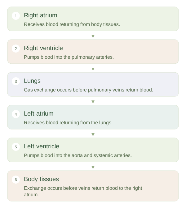
The heart is a muscular organ with right and left sides, each containing an above a . The right side receives blood returning from the body and sends it to the lungs. The left side receives blood from the lungs and sends it around the body. These are connected circuits: blood pumped by one side eventually reaches the other. Four chambers are useful because they separate receiving from forceful ejection and keep oxygen-poor and oxygen-rich blood largely separate in the normal adult circulation.
Four chambers, two connected circuits
 Open diagram larger ↗
Receiving and ejecting
Atria are relatively thin-walled receiving chambers. The right receives systemic venous blood, while the left atrium receives pulmonary venous blood. Much ventricular filling occurs while the atria and ventricles are relaxed, and atrial contraction adds the final contribution under ordinary conditions. Ventricles then generate pressure to eject blood into arteries. Valves between chambers and at ventricular exits guide one-way flow. The muscular separates the ventricular cavities, allowing the two sides to work at different pressures while normally pumping similar average volumes over time.
Why the left wall is thicker
The left has a thicker muscular wall because the systemic circuit generally requires greater pressure than the pulmonary circuit. The right ventricle pumps through the shorter, lower-resistance lung circulation. A thicker wall therefore reflects pressure work, not a need to pump a permanently larger blood volume than the right side. If one side consistently pumped more volume than the other, blood would accumulate somewhere in the circuit. The chambers also change shape and pressure throughout each beat. A diagram is a simplified map, so follow the connections rather than assuming the heart’s real orientation is a flat arrangement of four equal boxes.