Three protective coverings
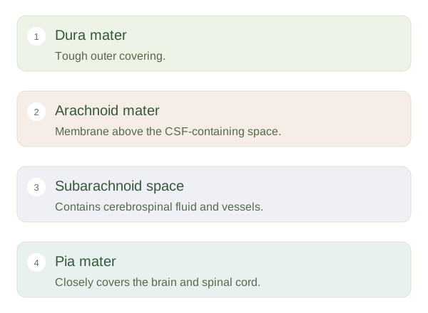
The brain and spinal cord are wrapped in three . The outer dura mater is tough and provides structural protection. The arachnoid mater lies beneath it, while the delicate pia mater closely follows the nervous tissue surface. Between arachnoid and pia is the , which contains , or CSF, and blood vessels. These layers help organise protection and support; they are not parts of the brain’s thinking cortex.
Meninges and cerebrospinal fluid protect the brain without making it immovable.
 Open diagram larger ↗
A circulating fluid cushion
CSF is produced mainly by choroid plexuses within the brain’s ventricles. It circulates through connected ventricular spaces and into the around the brain and spinal cord. It is returned through several drainage routes, including pathways associated with venous blood and lymphatic drainage. CSF provides buoyancy and cushioning, reducing the effective weight borne by supporting structures. It also contributes to a controlled environment and waste-clearance processes. The brain is protected, but can still be injured by sufficiently strong acceleration or pressure changes.
Location explains consequences
Inflammation of the is called meningitis. Blocked CSF circulation or impaired handling can contribute to hydrocephalus, enlargement of the ventricular system that may increase pressure. Bleeding in different meningeal compartments has different patterns because membranes and vessels are arranged differently. In the lower back, the spinal cord usually ends above the level used for a lumbar puncture. A needle can enter lumbar subarachnoid CSF below the cord, among mobile nerve roots. That anatomical relationship explains the procedure’s location without implying it is risk-free or suitable for untrained performance. Thinking about layers, fluid routes and limited space within the skull helps connect protective anatomy with why infection, bleeding or obstructed fluid circulation can become serious.