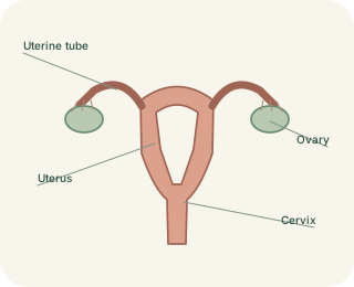
Ovaries and the released oocyte
The ovaries are paired organs in the pelvis. They contain follicles, each associated with an immature egg cell called an , and produce hormones including oestrogens and progesterone. Ovulation releases an oocyte from a mature follicle. The ovary is not directly joined to the open end of its uterine tube by a sealed pipe. Finger-like help guide the released oocyte towards the tube's opening.
From ovulation to the uterine cavity
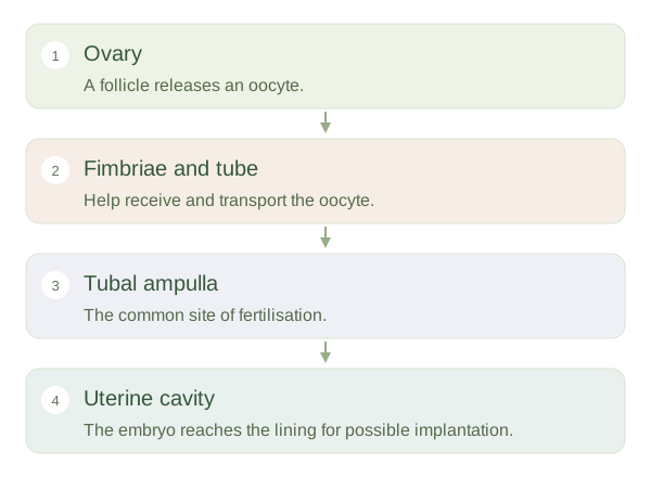 Open diagram larger ↗
A journey through the tube
The uterine tubes, also called fallopian tubes, extend from near the ovaries towards the uterus. Ciliated lining cells and smooth muscle activity help transport the or early embryo. Fertilisation most commonly occurs in the wider ampulla of a tube, rather than inside the uterus itself. The tube then provides a route towards the uterine cavity. A disrupted tube can affect fertility and can allow an embryo to implant outside the normal uterine cavity, producing an ectopic pregnancy.
The uterine wall
The uterus is a muscular organ located between the bladder and rectum in a common pelvic arrangement. Its inner changes across the menstrual cycle and can receive an implanting embryo. Its thick contains smooth muscle, important during birth. The lower cervix opens into the vagina, a muscular canal connecting to the exterior. Cervical mucus changes with hormonal conditions, influencing sperm passage. The vagina and urethra have separate openings; urine does not normally pass through the vagina.
A changing, supported system
Ligaments, connective tissue and pelvic-floor muscles help support these organs, but their exact positions vary with body shape, bladder filling, pregnancy and other factors. The system is dynamic: a follicle grows, an is released, and the uterine lining changes in response to hormones. Distinguishing these tasks helps explain why producing an egg, fertilisation and implantation are separate events in separate locations, rather than one event occurring entirely within the uterus.