A fluid-filled sound analyser
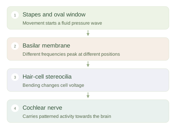
The is a coiled structure in the inner ear. Movement of the stapes at the oval window creates pressure waves in its fluid, while the round window permits pressure displacement. Inside, the supports the organ of Corti, which contains sensory hair cells. These cells do not detect air directly. They respond to the relative movement of nearby structures as a wave travels through the cochlea. Their microscopic projections are called stereocilia, despite not behaving like the actively beating cilia of the airways.
From vibration to a hearing signal
 Open diagram larger ↗
Different places, different pitches
The varies in stiffness and width. Its base, near the oval window, responds best to high frequencies, while regions nearer the apex respond best to lower frequencies. A complex sound activates a pattern along the membrane, allowing the nervous system to distinguish its components. This spatial frequency arrangement is called . Loudness is represented partly by the strength and spread of neural activity, rather than by one special location. Pitch and loudness are therefore related to different features of the cochlear response.
From bending to hearing
Bending stereocilia changes the opening of mechanically sensitive channels, altering the hair cell’s voltage and transmitter release onto nerve fibres. Inner hair cells supply most of the information sent through the cochlear nerve. Outer hair cells change length and boost local membrane motion, sharpening sensitivity and frequency selection. Signals pass through several brainstem stations and onwards to auditory cortex, where meaningful sound is perceived. Damage to hair cells can reduce hearing even if sound transmission through the outer and middle ear is normal. Hearing depends on a mechanical stage, a sensory stage and neural interpretation working together.