Protection with planned connections
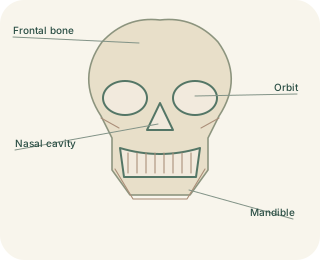
The skull forms a protective case for the brain and a framework for the face. Its cranial bones include the frontal, parietal, temporal, occipital, sphenoid and ethmoid bones. Most meet at fibrous joints called sutures. These are not normal freely moving joints, but they allow growth during development before becoming more tightly united. Facial bones form structures such as the upper jaw, cheekbones and much of the nasal framework. The is the lower jaw and moves at the temporomandibular joints.
Important skull passageways
 Open diagram larger ↗
Openings are functional routes
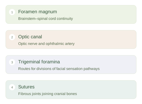
A protected brain still needs nerves and blood vessels to communicate with the rest of the body. Openings called foramina provide these routes. The magnum in the occipital bone permits continuity between the brainstem and spinal cord, alongside associated structures. The optic canal carries the optic nerve and ophthalmic artery towards the orbit. Other foramina transmit branches of the trigeminal nerve or vessels supplying the head. An opening is therefore not simply a weakness: it is a necessary passage through a rigid boundary.
Reading the skull as a map
The cranial floor has anterior, middle and posterior fossae, broad depressions supporting different parts of the brain. Their relationships help explain why injury near one opening may affect a particular nerve. The temporal bones contain delicate hearing and balance structures, while the ethmoid contributes to the roof of the nasal cavity. Some skull bones contain air-filled sinuses, but they are not hollow substitutes for the entire skull. Learning landmarks and the structures passing through them is more useful initially than memorising every small hole. The central concept is a strong framework that protects organs while preserving essential connections.