A muscular partition
The diaphragm separates the thoracic and abdominal cavities. Its muscle fibres attach around the lower thoracic opening and converge towards a . At rest it forms domes, with the right usually higher because of the liver beneath it. When its fibres contract, the domes descend and thoracic volume increases. This helps lower pressure within the chest so air flows into the lungs. The diaphragm does not pull air through a tube itself; it changes the space around the lungs, allowing pressure differences to drive airflow.
Main routes across the diaphragm
 Open diagram larger ↗
Crossing the partition
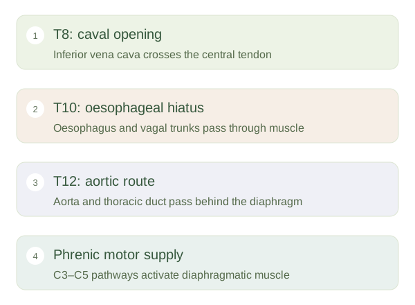
Important structures must travel between chest and abdomen without interrupting normal movement. The inferior vena cava passes through an opening in the , commonly described at vertebral level T8. The oesophagus passes through a muscular opening at about T10, accompanied by vagal trunks. The aorta passes behind the diaphragm beneath a ligamentous arch around T12, alongside the . These levels are useful approximate landmarks, not fixed measurements for every body position. The aortic route is arranged so diaphragmatic contraction does not normally compress the main artery as it descends.
Control and wider effects
The phrenic nerves, arising chiefly from C3 to C5 spinal levels, provide motor supply to the diaphragm. Their long course links the neck with the chest. Diaphragmatic movement also changes abdominal pressure and helps coordinate activities such as coughing and straining. During quiet expiration, relaxation allows elastic recoil to reduce lung volume; the diaphragm need not actively push air out on every normal breath. Its dual identity as both breathing muscle and anatomical partition explains why its openings, nerve supply and relationships are important across respiratory, digestive and regional anatomy.