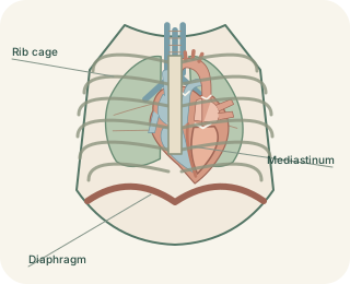
The region between the lungs
The is the central thoracic region between the two pleural sacs. It extends from the above to the diaphragm below, with the sternum in front and thoracic vertebral column behind. It contains the heart, major vessels, airway and food passage, nerves, lymphatic structures and connective tissue. The lungs themselves are lateral to it within their pleural spaces. This distinction makes a chest image easier to interpret: an opacity in the central compartment is not automatically located in lung tissue.
Traditional mediastinal compartments
 Open diagram larger ↗
A useful anatomical division
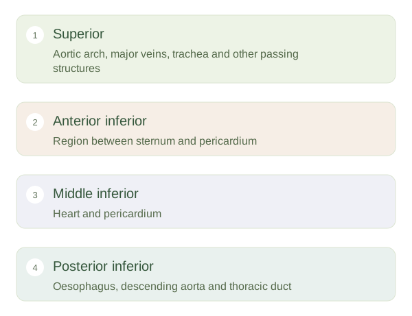
A plane through the towards the T4–T5 region separates superior from inferior in the traditional anatomical model. The superior part includes structures such as the aortic arch, major veins, trachea and oesophagus. The inferior mediastinum is divided into anterior, middle and posterior parts. The middle contains the heart and . The anterior part is a relatively small region between the sternum and pericardium, while the posterior part lies behind the pericardium and includes the descending thoracic aorta, oesophagus and thoracic duct.
Connections matter more than boxes
These divisions are descriptive regions rather than separate sealed chambers. The oesophagus, vessels and nerves cross boundaries as they travel between neck, chest and abdomen. Phrenic nerves descend towards the diaphragm, while vagal pathways contribute to organ control. The thymus is more prominent earlier in life and often becomes less conspicuous as fatty tissue increases with age. Imaging specialists may use compartment models that differ from the traditional anatomical divisions. The practical aim is to understand neighbouring structures and continuous routes, rather than assuming every chest description uses exactly the same set of borders.