Two lungs with different outlines
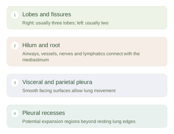
The right lung usually has three lobes, separated by horizontal and oblique fissures. The left usually has two lobes separated by an oblique and includes a cardiac notch along its anterior border. Its lingula belongs to the upper lobe, rather than being a separate third lobe. Each lobe contains smaller bronchopulmonary segments, supplied by branching airways and vessels. These divisions help organise the organ, but gas exchange still depends on the microscopic alveolar surfaces distributed within them.
Lung tissue, connections and surrounding space
 Open diagram larger ↗
A doorway on the medial surface
The is the region on the lung’s medial surface where major structures enter or leave. The root is the collection of structures passing through it, including a main bronchus, pulmonary vessels, bronchial vessels, lymphatics and nerves. Pulmonary arteries carry blood towards gas exchange, while pulmonary veins return blood towards the left atrium. Bronchial vessels serve the supporting tissues of the lung itself. The hilum is therefore a connected doorway, not an air-filled room. Its contents link the lung with central thoracic structures in the mediastinum.
Pleural reflections and spare room
covers the lung surface, while parietal pleura lines the chest wall, diaphragm and mediastinal boundary. A thin lubricated space normally lies between them. Where parietal surfaces meet beyond the resting lung margins, pleural recesses provide room for expansion. The costodiaphragmatic recess lies near the lower chest between costal and diaphragmatic pleura. Fluid can collect in dependent pleural regions, and lung edges move with breathing. A simple outline on a diagram therefore represents one moment in a moving system. Understanding the difference between lung tissue, hilar structures and pleural space helps avoid confusing nearby but functionally distinct compartments.