Cord inside a protected canal
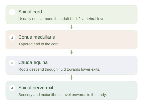
The spinal canal is formed by successive vertebral openings. Within it, the spinal cord is surrounded by protective membranes called and by cerebrospinal fluid. The cord contains pathways carrying information between the brain and body, as well as circuits involved in reflexes. It does not extend to the lowest end of the adult vertebral column. Its tapered end, the , is usually around the L1–L2 vertebral level, with individual variation. This difference between the length of the cord and the canal is central to understanding lower-back anatomy.
Spinal Canal, Nerve Roots and Cauda Equina — concept map
 Open diagram larger ↗
Roots travel to their exits
At each spinal cord segment, sensory fibres enter through dorsal roots and motor fibres leave through ventral roots. These roots join to form mixed spinal nerves. As the body grows, the vertebral column becomes longer relative to the cord. Roots destined for lower exits therefore descend inside the canal before leaving. Below the cord they form a bundle resembling a horse’s tail, called the . These are peripheral nerve roots rather than a continuation of solid spinal cord tissue. Their route means a problem inside the lower canal can affect roots travelling towards several different exits.
Levels do not always match
A vertebra, a spinal cord segment and a spinal nerve are related labels, but they are not interchangeable. Lower cord segments lie above the similarly numbered vertebrae because their roots travel downwards. Clinicians use both imaging and patterns of nerve function to localise a problem. The meningeal sac continues below the cord, leaving a fluid-filled lower region containing nerve roots. This explains the anatomical basis for accessing cerebrospinal fluid in the lumbar region, although the procedure requires professional technique. The key idea is that the contents of the canal change along its length: upper levels contain cord, while lower levels chiefly contain descending roots.