A muscular floor with openings
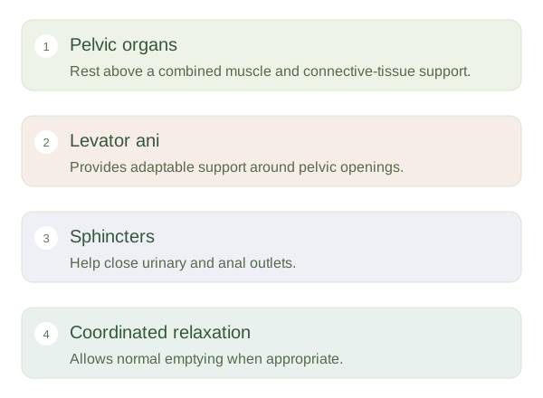
The pelvic floor closes much of the bottom of the pelvic cavity. Its major muscular component is the , formed chiefly by levator ani and coccygeus. Connective tissues link the muscles to the pelvic walls and to nearby organs. Openings allow the urethra, anal canal and, when present, vagina to pass through. This means the floor cannot simply be a solid sheet. It must support the contents above while allowing urine, stool and, in childbirth, a baby to pass through appropriately controlled routes.
Pelvic Floor and Continence — concept map
 Open diagram larger ↗
Continence is a team function
means retaining urine or stool until release is appropriate. Pelvic floor muscles work with urethral and anal sphincters, organ walls, connective tissues and the nervous system. Part of levator ani, , forms a sling around the anorectal junction and helps maintain its angle. The external anal sphincter provides additional voluntary control. During a cough, abdominal pressure rises quickly; coordinated pelvic floor and sphincter responses help prevent leakage. During normal emptying, some of these muscles must relax rather than keep tightening. Effective function therefore involves timing and relaxation as well as force.
Support across changing demands
Pregnancy, childbirth, ageing, pelvic surgery, tissue characteristics and long-term pressure changes can affect the supporting system. None predicts the same outcome for every person. Problems can occur when muscles are weak, poorly coordinated or excessively tense. This is why all pelvic symptoms cannot be reduced to a need for stronger muscles. Nerves provide both sensation and control, allowing reflex responses and deliberate participation. The pelvic floor also works with the diaphragm and abdominal wall as pressure changes during breathing and movement. Its role is dynamic: it supports, responds and releases according to the task, rather than holding the pelvic outlets permanently closed.