Below the pelvic diaphragm
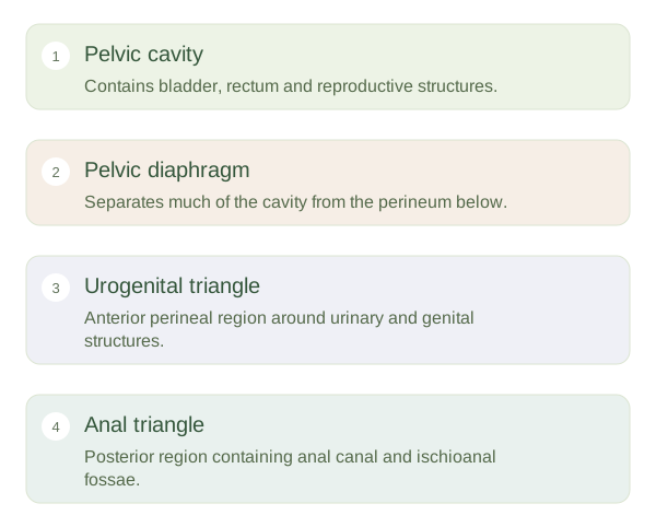
The is the region below the pelvic diaphragm and between the upper thighs. Its bony landmarks form a diamond: the pubic symphysis in front, the coccyx behind, and the ischial tuberosities at the sides. A line between the ischial tuberosities divides the diamond into an anterior and a posterior anal triangle. This is a map of the region, not a physical partition running through the skin. The perineum is different from the whole pelvic cavity, which lies above the floor and contains organs such as the bladder and rectum.
Perineum and Pelvic Compartments — concept map
 Open diagram larger ↗
Outlets and supporting tissues
The contains external genital structures and tissues associated with the urinary outlet. Its detailed arrangement differs according to the person’s anatomy. The perineal membrane provides support in the anterior region. The anal triangle contains the anal canal, external anal sphincter and fat-filled ischioanal fossae on either side. This fat allows the canal to expand during defaecation while cushioning nearby structures. The is a central fibromuscular meeting point where several supporting structures attach. It is especially important to the integrity of the region around the vaginal and anal openings.
Compartments guide understanding
The pelvis itself can be described using anterior urinary, middle reproductive and posterior bowel relationships, although these structures remain closely connected through nerves, vessels and supporting tissues. Perineal spaces describe tissue planes below the pelvic floor rather than empty rooms. These planes influence how swelling or infection can spread. The pudendal nerve supplies much of the perineal skin and provides motor control to important voluntary sphincter and perineal muscles. Recognising these layers prevents a common confusion: an organ can lie in the pelvis while its outlet passes through the . Location, support and nerve supply must be considered together to understand the region.