A hinge with extra movement
The knee joins the femur to the tibia and also includes the patellofemoral articulation. It mainly bends and straightens, but small rotations accompany movement. Rounded femoral condyles meet relatively shallow tibial surfaces, so bone shape alone provides limited stability. Articular cartilage covers the contacting surfaces to reduce friction and spread load. The sits within the quadriceps tendon and improves the muscle’s leverage. It contacts the femur, rather than forming a direct joint with the tibia.
Different jobs at the knee
 Open diagram larger ↗
Load sharing and restraint
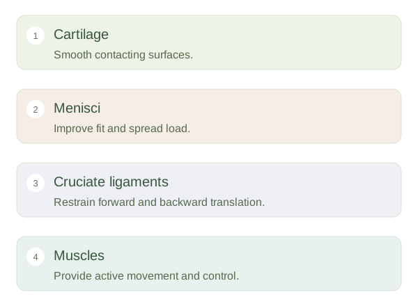
Two crescent-shaped menisci of fibrocartilage sit between femur and tibia. They improve the fit of the surfaces and distribute weight over a larger area. The anterior limits forward movement of the tibia relative to the femur; the posterior cruciate ligament limits backward movement. The collateral ligaments resist excessive sideways angulation. These structures also contribute to rotational control. Their names describe position or course, but their function is easier to remember by imagining which unwanted movement would tighten them.
Movement changes the demands
During walking, muscles control how quickly the knee bends and straightens while ligaments guide its limits. A planted foot combined with sudden rotation creates different stresses from a slow straight-line bend. Injury can therefore involve more than one structure. Menisci have an uneven blood supply, with better vascular access near their outer edges, so healing potential varies by tear location. Pain or swelling does not reveal a precise injured structure without further assessment. The useful concept is shared responsibility: cartilage handles smooth contact, menisci improve load distribution and ligaments restrain displacement, while muscles provide active control.