What anatomists mean by leg
In everyday speech, leg may mean the whole lower limb. Anatomically, the leg is the region between knee and ankle. The tibia bears most of the weight, while the fibula provides muscle attachments and contributes to ankle stability. Deep fascia and the membrane between these bones divide muscles into anterior, lateral and posterior compartments. The posterior region has superficial and deep groups. Compartments contain nerves and blood vessels as well as muscles, so their boundaries are important functional relationships, not just convenient labels on a diagram.
Leg movement map
 Open diagram larger ↗
A movement map
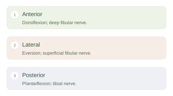
Anterior muscles, including tibialis anterior, lift the forefoot in and help extend the toes. Their main nerve is the deep fibular nerve. Lateral fibularis muscles help evert the foot, turning its sole outwards, and receive the superficial fibular nerve. Posterior muscles mainly point the foot down in and help flex the toes. Gastrocnemius and soleus form the calf and transmit force through the calcaneal tendon. The tibial nerve supplies the posterior compartments. Some muscles also invert the foot, so compartments describe dominant patterns rather than a single shared action.
Space and pressure matter
Fascia does not expand easily. Marked swelling within a compartment can raise pressure enough to impair blood flow and nerve function. This explains why compartment syndrome is a problem of tissue perfusion, not simply a large bruise. The anatomical map also helps explain foot drop: weakness of makes it harder to clear the toes during swing. A nerve problem can occur at different levels, however, so the movement finding alone does not prove one particular lesion. Structure, movement, sensation and the wider history must be considered together.