Cells within supportive matrix
Cartilage and bone are connective tissues whose cells occupy spaces called lacunae within a substantial matrix. Cartilage cells are chondrocytes; bone-maintaining cells are osteocytes. This similarity can make the tissues seem interchangeable, but their matrix and nutrient supply differ greatly. Cartilage commonly has a firm, hydrated matrix that resists compression while retaining some flexibility. Bone combines collagen with mineral, making a strong composite capable of supporting greater rigid loads. Their microscopic patterns reflect the different jobs each tissue performs in the skeleton.
Two supportive tissues
 Open diagram larger ↗
Cartilage is not one uniform tissue
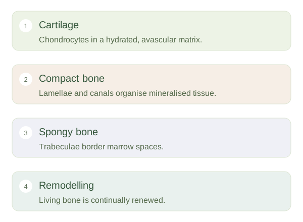
Hyaline cartilage has a relatively smooth-looking matrix on routine stains and occurs at many articular surfaces and in the airways. Elastic cartilage contains additional elastic fibres, as in parts of the outer ear. Fibrocartilage contains prominent collagen bundles and occurs in structures such as intervertebral discs. Cartilage lacks blood vessels within its matrix, so cells receive nutrients mainly by diffusion. Some cartilage has a surrounding perichondrium, but articular cartilage does not. These supply limitations help explain why cartilage repair can be slow or incomplete.
Bone organises around supply
Compact bone often contains osteons: concentric lamellae arranged around central canals carrying vessels and nerves. Tiny canaliculi connect osteocyte spaces, helping exchange signals and substances. Spongy bone forms a network of trabeculae with marrow spaces rather than dense collections of complete osteons. Osteoblasts form new matrix and osteoclasts remove bone during remodelling. Mineral does not make bone dead or unchanging; it remains metabolically active tissue. When inspecting a slide, look for matrix arrangement, vascular spaces and cell organisation, then connect those features to load support, maintenance and the ability to respond to changing forces.