From a cell to organised layers
After fertilisation, the developing conceptus divides repeatedly and later implants in the uterine lining. Early development is not just enlargement: cells adopt different roles and positions. During , three major germ layers form. Ectoderm contributes to the nervous system and outer skin layer. Mesoderm contributes to tissues including muscle, bone, blood and much of the cardiovascular system. Endoderm contributes much of the epithelial lining of the digestive and respiratory tracts. Each organ usually combines contributions from more than one layer rather than developing from a single uniform material.
The Embryonic Body Plan
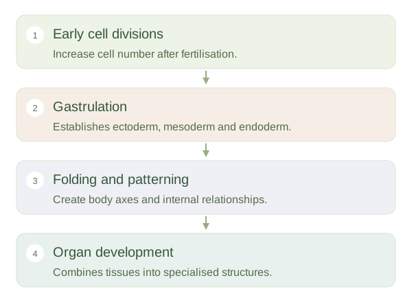 Open diagram larger ↗
Folding creates relationships
The early embryonic disc changes shape through coordinated folding. This helps enclose a primitive gut and establishes the relationship between the body wall and internal structures. A neural plate folds into a , which gives rise to the brain and spinal cord. Cells called neural crest cells separate from its edges and contribute to diverse structures, including much of the peripheral nervous system. The also establishes head–tail, back–front and right–left organisation. These axes provide the framework within which organs take shape and connect with one another.
Development explains adult anatomy
Some adult relationships make more sense when their developmental history is considered. The heart and gut change position and shape as the grows, while repeated tissue segments contribute to the organisation of the trunk. Development is controlled by interacting genetic signals and physical processes; it is not a miniature adult simply unfolding. Timing matters because different structures are forming at different stages. A disturbance can therefore affect a particular set of structures during a sensitive window. These principles provide a foundation for understanding congenital differences without assuming that every developmental variation is harmful or that one gene determines every feature.