One organ, several regions
A typical long bone has a shaft, called the , and expanded ends, called epiphyses. Between them are transition regions associated with growth during childhood. The outside is covered by except where articular cartilage covers joint surfaces. The periosteum contains vessels, nerves and cells important for repair and growth in width. Inside, an endosteal lining covers many internal bone surfaces. These coverings are living interfaces, not decorative wrappers. Blood vessels enter through openings and supply both the bone tissue and marrow within.
Inside a Bone
 Open diagram larger ↗
Dense shell and internal network
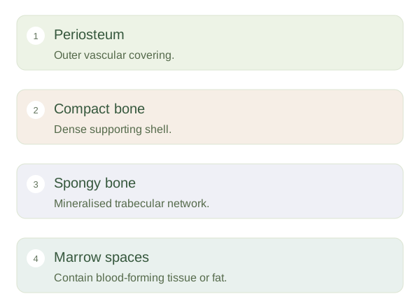
Compact bone forms a dense outer layer, especially thick along the shaft. Much of it is organised into cylindrical units called osteons, with matrix arranged around small channels containing vessels and nerves. Spongy bone contains a network of trabeculae with spaces between them. The trabeculae help distribute loads and are oriented in relation to mechanical demands. Spongy does not mean soft or unimportant: the tissue is mineralised, but its arrangement leaves space for marrow and reduces the mass needed to support a given load.
Cells maintain the structure
Osteoblasts build new matrix, osteoclasts resorb bone and osteocytes maintain and sense conditions within the matrix. These activities are coordinated rather than simply competing at random. A marrow cavity occupies much of the shaft of a long bone, while marrow also fills spaces among trabeculae. Near joint surfaces, smooth articular cartilage reduces friction and distributes pressure. No single drawing represents every bone: flat bones, for example, have different shapes and arrangements. Nevertheless, the same principles recur—vascular living tissue, a composite matrix and architecture matched to function. A cross-section is useful because it reveals this organisation that an external view hides.