A firm but flexible tissue
Cartilage is connective tissue containing cells called chondrocytes within a water-rich extracellular matrix. Its collagen and other components allow it to resist compression while retaining some flexibility. Unlike most bone, cartilage normally lacks its own blood vessels. Nutrients must diffuse through the matrix from surrounding sources. This helps explain why damaged cartilage often repairs slowly or incompletely. It is not simply immature bone: although some bone develops by replacing cartilage, many cartilage structures normally remain throughout life and perform specialised jobs.
Cartilage and Its Functions
 Open diagram larger ↗
Three useful patterns
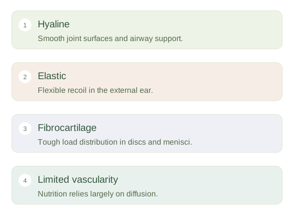
provides smooth articular surfaces at many joints and supports structures such as the trachea and costal cartilages. Its matrix is suited to low-friction movement and compressive loading. contains abundant elastic fibres, allowing flexible structures such as the outer ear to spring back after bending. has strong collagen bundles and is found in load-bearing structures such as intervertebral discs and knee menisci. These categories describe differences in matrix organisation. They help explain why an ear can fold while a joint surface needs to remain smooth under repeated loading.
Working at a joint
Articular cartilage distributes forces across the ends of bones. Its water-rich matrix deforms under load and recovers when the load changes. Synovial fluid lubricates the surface and helps nourish the tissue. Repeated normal movement assists exchange and load distribution, but excessive or abnormal forces can damage the surface. Once cartilage is lost, the underlying bone does not become an equivalent smooth replacement. Joint disease therefore involves altered mechanics as well as tissue changes. When studying cartilage, connect its location to its matrix: smooth support, elastic recoil and resistance to heavy combined loading require different structural solutions.