A moving interface
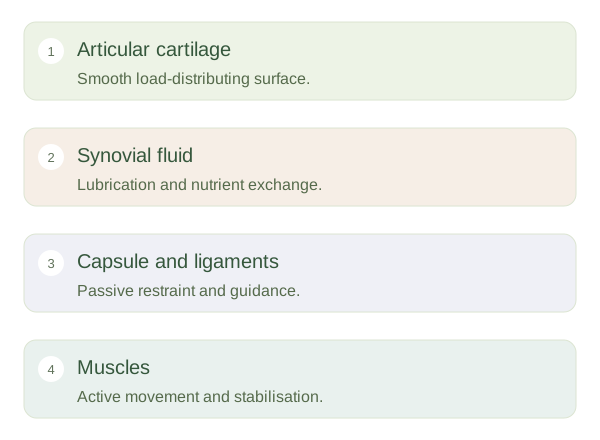
In a synovial joint, bone ends are separated by a narrow joint cavity. Their articular surfaces are usually covered by hyaline cartilage, creating smooth contact regions that distribute pressure. Cartilage is not the same as the bone beneath it: its water-rich matrix deforms and recovers under load. A surrounding encloses the cavity. Its outer fibrous layer provides restraint and continuity with nearby connective tissues, while an inner lines internal surfaces except the articular cartilage and certain other specialised surfaces.
Inside a Synovial Joint
 Open diagram larger ↗
Fluid and supporting structures
The contributes to , which lubricates the moving surfaces and helps nourish avascular articular cartilage. A small fluid volume is normal; a large effusion is different from simply having a joint cavity. Ligaments reinforce and guide the joint, and nearby muscles provide dynamic control. Some joints include additional fibrocartilage structures, such as menisci, that improve fit or distribute load. Bursae are fluid-containing sacs that reduce friction between nearby moving tissues, but they are not identical to the main articular cavity.
A coordinated mechanical system
Joint stability depends on the fit of its surfaces, passive restraints and active muscle control. These contributions differ by joint. A deep hip socket provides substantial bony containment, while the shoulder relies more heavily on soft tissues for its wide mobility. Movement also helps distribute across cartilage surfaces. Pain or swelling can change muscle activation and movement patterns, so injury to one component affects the system as a whole. When examining a joint diagram, identify what bears load, what lubricates, what restrains excess movement and what actively moves the bones. Each component solves a different part of the same mechanical problem.