Bundles within bundles
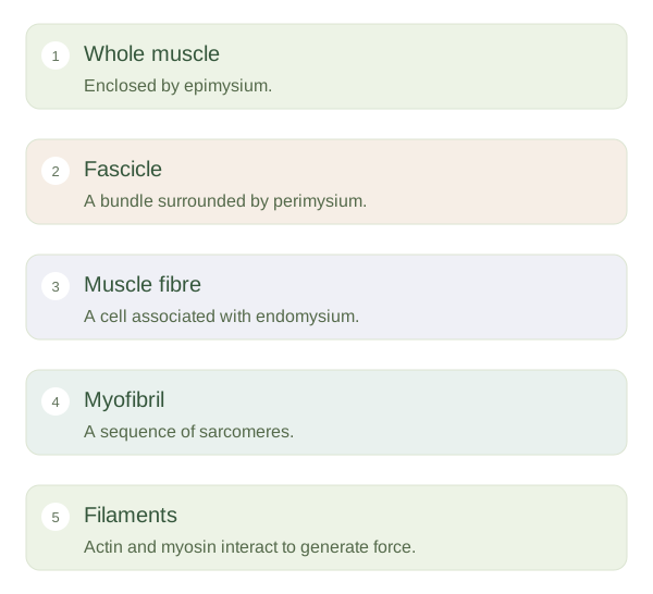
A skeletal muscle is an organ containing muscle fibres, connective tissues, blood vessels and nerves. Fibres are grouped into bundles called fascicles. Epimysium surrounds the whole muscle, perimysium surrounds fascicles and endomysium surrounds individual fibres. These layers connect with one another and transmit force towards tendons or other attachments. They also provide routes for vessels and nerves. The arrangement resembles organised cables within a larger cable, although living muscle is far more adaptable and contains active cells rather than passive wires.
Inside a Skeletal Muscle
 Open diagram larger ↗
Inside one fibre
A is a long cell containing many nuclei. Its membrane is called the sarcolemma, and inward membrane extensions called transverse tubules carry electrical changes into the cell. The sarcoplasmic reticulum stores calcium needed to activate contraction. Most of the fibre’s interior is occupied by myofibrils, long structures made of repeating sarcomeres. A extends between two Z discs and contains thin actin filaments and thick myosin filaments. Their regular arrangement produces the striped appearance seen under a microscope. Individual filaments slide rather than shrinking during normal contraction.
Organisation supports function
When many sarcomeres shorten in sequence, a can shorten substantially. Arranging myofibrils side by side allows their forces to add together. This helps explain why muscle architecture influences performance: length, cross-sectional area and fibre direction all matter. A long fibre can contribute to a large shortening distance, while many fibres acting in parallel can increase force capacity. Nerve input, calcium handling and energy supply must support this machinery. When reading a diagram, keep the levels separate: a fibre is a whole cell, a myofibril is a structure inside that cell and a is a repeating unit within a myofibril.