The signal reaches the fibre
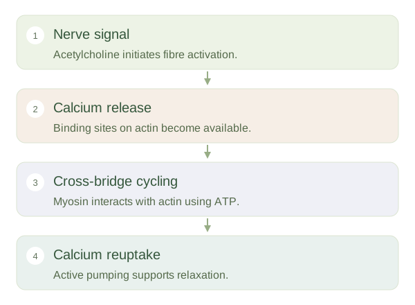
A motor neuron releases acetylcholine at the . This chemical activates receptors on the muscle membrane, initiating an electrical response when the stimulus is sufficient. An action potential travels along the sarcolemma and into transverse tubules. The electrical change triggers calcium release from the . This arrangement helps activate the fibre’s interior rapidly rather than relying on a slow signal diffusing from its outer surface. The nerve signal begins the process, but the force itself is generated by interactions between proteins inside the muscle cell.
How Muscle Fibres Contract
 Open diagram larger ↗
Filaments slide
Calcium binds to , changing the position of tropomyosin so binding sites on actin become accessible. Myosin heads attach to actin and undergo a force-producing movement. ATP binding allows myosin to detach, and ATP breakdown prepares the head for another cycle. Repeated interactions pull thin filaments further between thick filaments, shortening the sarcomere when the load permits. The actin and myosin filaments themselves do not become shorter. Force can also develop without overall muscle shortening, as when holding a heavy object still against gravity.
Relaxation is active too
When neural stimulation stops, acetylcholine is cleared and calcium is pumped back into its storage compartment. This pumping uses energy. As calcium falls, the regulatory proteins again limit actin–myosin binding. ATP is also needed for myosin detachment, so relaxation is not simply what happens when energy disappears. A functioning muscle needs both suitable activation and an ongoing energy supply. The same basic sequence can be varied in timing and recruitment to produce a gentle movement or a powerful effort. Remember the causal order: electrical activation changes calcium availability, calcium permits interaction, and ATP supports the molecular cycle.