Four one-way gateways
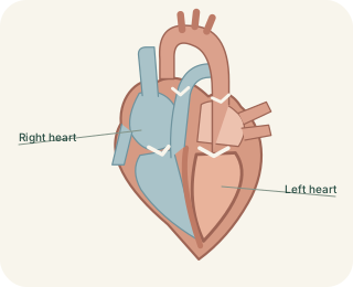
The heart has two atrioventricular valves and two semilunar valves. The tricuspid valve lies between the right atrium and right ventricle, while the lies between the left atrium and left ventricle. The pulmonary valve guards the right ventricular outlet and the aortic valve guards the left ventricular outlet. Their leaflets are flexible structures attached to supporting tissues. The valves do not actively pull themselves open like a powered door: changing pressure on either side determines whether their leaflets separate or come together.
Heart Valves and One-Way Flow
 Open diagram larger ↗
Pressure drives movement
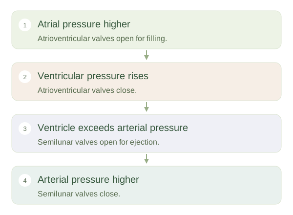
During ventricular filling, atrial pressure exceeds ventricular pressure enough to open the atrioventricular valves. When ventricular contraction raises pressure above atrial pressure, those valves close. Once ventricular pressure exceeds the pressure in the receiving artery, the corresponding semilunar valve opens. As the ventricle relaxes and arterial pressure becomes greater, a brief tendency towards reverse flow fills the semilunar pockets and closes them. The first main heart sound is associated with atrioventricular valve closure; the second is associated with semilunar closure. The sounds involve vibrations of the valve–blood–heart system, not simply doors clicking.
Supporting the leaflets
link atrioventricular valve leaflets to papillary muscles within the ventricles. When the ventricles contract, papillary muscle tension helps prevent the leaflets from prolapsing into the atria. It does not normally pull the valves open. Semilunar valves do not use the same chordal arrangement. If a valve narrows, called , forward flow faces extra resistance. If it does not close effectively, allows backward flow. These mechanisms change the work required of the chambers. Understanding valves means following pressure and direction, rather than memorising that one valve opens whenever another happens to close.