Returning from the body
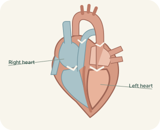
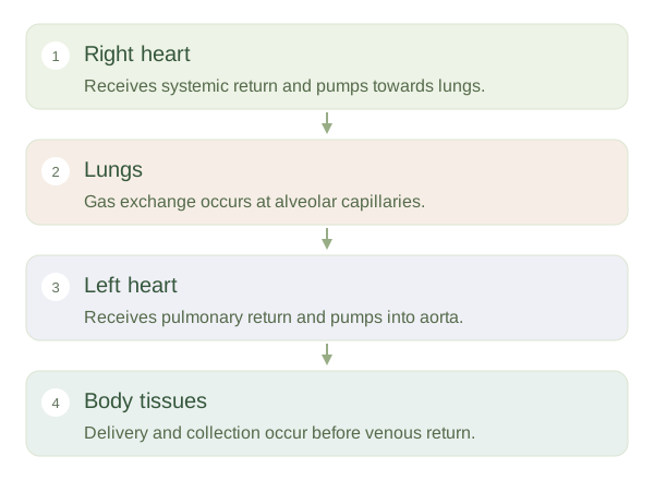
After supplying systemic tissues, blood returns through veins towards the right atrium. The superior vena cava drains much of the upper body, and the inferior vena cava returns blood from below the diaphragm. Coronary venous blood also reaches the right atrium. This returning blood still contains oxygen, but generally less than blood leaving the lungs. It passes through the tricuspid valve into the right ventricle. When that ventricle contracts, blood travels through the pulmonary valve into the pulmonary trunk and pulmonary arteries.
The Route of Blood Through the Body
 Open diagram larger ↗
Through lungs to the left heart
Pulmonary arteries carry blood to the lungs, where small vessels form networks around alveoli. Oxygen moves into blood and carbon dioxide moves towards the alveolar air according to their partial-pressure differences. Pulmonary veins then return blood to the left atrium. It passes through the mitral valve into the left ventricle, which ejects it through the aortic valve into the aorta. The names artery and vein describe direction relative to the heart, not oxygen content. Pulmonary arteries and veins are therefore useful reminders that the usual systemic oxygen pattern has exceptions.
Delivery and exchange
The aorta branches into systemic arteries, smaller arterioles and capillaries. At capillary beds, gases, nutrients, water and other substances exchange with surrounding tissues. Blood then collects into venules and veins to complete the circuit. Many systemic organs receive blood through parallel branches, allowing flow to be distributed according to need; blood does not normally visit every organ in a single fixed sequence. Some specialised arrangements, such as the hepatic portal circulation, add another capillary bed before blood returns to the heart. The overall two-circuit map remains the starting framework. Trace it by direction and connections rather than by red and blue colours, which are diagram conventions rather than the actual colours of oxygenated and deoxygenated blood.