A rhythm begins inside the heart
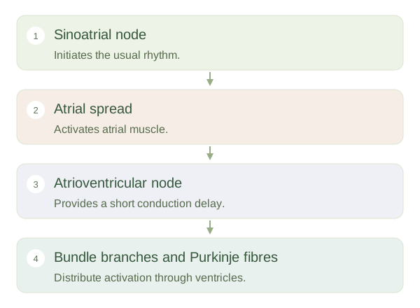
Specialised cardiac cells can spontaneously generate electrical activity. Under normal conditions, the in the right atrium acts as the main pacemaker because its rhythm sets the pace for the rest of the heart. The electrical signal spreads through atrial muscle, supporting coordinated atrial contraction. The autonomic nervous system and circulating hormones adjust the rate and conduction, but the heart does not need a separate motor-nerve command for every beat. This intrinsic rhythm is a major difference from ordinary skeletal muscle activation.
The Heart’s Electrical System
 Open diagram larger ↗
A useful pause and rapid spread
The signal reaches the , where conduction slows. This delay allows the atria to finish their contribution to ventricular filling before the ventricles contract strongly. The signal then travels through the atrioventricular bundle, right and left bundle branches and Purkinje fibres. Rapid distribution through the ventricles helps activate the working muscle in a coordinated pattern, broadly promoting ejection from the ventricular regions towards the outflow tracts. The fibrous skeleton between atria and ventricles electrically insulates much of the boundary, making the normal conduction pathway important.
Electricity precedes mechanics
An electrocardiogram, or , records electrical voltage differences detected at the body surface. The P wave reflects atrial , the QRS complex ventricular depolarisation and the T wave ventricular repolarisation. These are electrical events, not direct measurements of pumping strength or blood flow. Mechanical contraction follows activation, so a recognisable electrical pattern does not by itself prove effective circulation. Changes in rhythm or conduction can disturb chamber timing and therefore filling or ejection. The central concept is coordination: a heartbeat needs not just active muscle cells, but activity delivered in a sequence that allows the connected chambers to function as pumps.