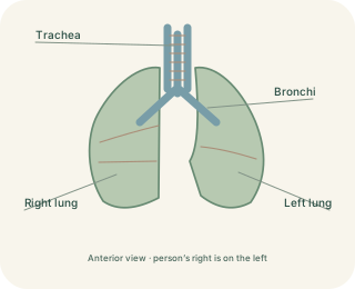
Two surfaces, a tiny space
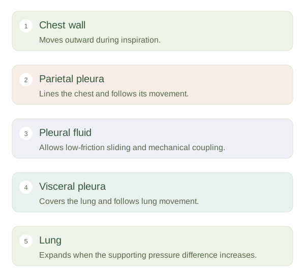
Each lung is covered by , a thin membrane closely attached to its surface. lines the inner chest wall, diaphragm and mediastinal boundary. Between them lies the pleural cavity, normally a very narrow potential space containing a small amount of fluid. A useful analogy is two wet glass slides: they can glide across one another, but separating them is harder. The analogy illustrates coupling, although living pleural tissues and pressures are more complex than glass slides.
Pleural layers let lungs slide while following the chest wall.
 Open diagram larger ↗
Why pressure matters
The lungs naturally recoil inward because their tissues are elastic and their moist air spaces generate surface tension. The chest wall has an opposing tendency at ordinary resting volumes. In the sealed pleural arrangement, this opposition creates a pressure that is normally below atmospheric pressure during quiet breathing. The pressure difference across the lung helps keep it expanded. When the diaphragm contracts and the chest enlarges, pleural pressure becomes more negative. The lung expands with the chest and air enters because alveolar pressure falls below outside air pressure.
When coupling is disturbed
If air enters a pleural cavity, it can reduce the pressure difference supporting that lung. This is a ; the lung may recoil partly or extensively. Fluid collecting in the pleural space is called a and may limit expansion. Neither normally belongs inside the alveoli, although different illnesses can affect several compartments together. The right and left pleural cavities are usually separate, which limits direct spread of a problem from one side to the other. Pleural fluid normally reduces friction during thousands of daily breaths. Understanding these membranes explains why chest movement, lung inflation and conditions around the lung are connected, even though the lung itself does not contain a muscle that actively pulls it open.