A long tube with an enormous surface
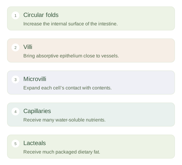
The small intestine consists of the duodenum, jejunum and ileum. Despite its name, it provides much of the digestive tract’s length and the main site for nutrient absorption. Circular folds increase internal surface area. Finger-like villi project from the mucosa, and the absorptive cells covering them have microscopic projections called . These are three different levels of surface enlargement. They allow many transport proteins and digestive surfaces to contact the intestinal contents within a limited abdominal space.
A folded surface and short transport distance make absorption efficient.
 Open diagram larger ↗
A living interface, not a simple sieve
The epithelial layer is thin, but substances do not all pass through freely. Transport proteins, enzymes and cell membranes determine which molecules cross and how. Some movement is passive, while other transport depends directly or indirectly on cellular energy. Brush-border enzymes on complete parts of digestion close to the absorptive surface. Tight junctions help regulate movement between cells and contribute to the barrier separating gut contents from internal tissues.
Where absorbed material goes
Each contains blood capillaries and a lymphatic vessel called a . Many sugars, amino acids and water-soluble substances enter blood that travels towards the liver through the portal circulation. Much absorbed dietary fat is packaged by intestinal cells and enters lacteals before reaching the bloodstream through lymphatic routes. The intestine must also balance absorption with immune defence and continuous renewal of its lining. Damage that flattens villi reduces functional surface area and can impair nutrient absorption even when the overall tube remains open. The key concept is that structure supports selective transfer: the folds increase contact, the thin lining shortens the crossing and separate blood and lymph routes carry different absorbed products onward.