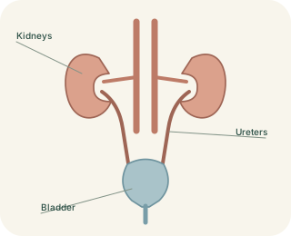
A protected position behind the abdomen
The kidneys lie towards the back of the abdomen, mainly behind the peritoneal lining. They are therefore described as . The right kidney is usually slightly lower than the left because of neighbouring anatomy, including the liver. Each kidney has an outer cortex and an inner medulla. The medulla contains pyramids whose tips drain into collecting spaces. Fat, fascia and a fibrous capsule help support and protect the organ without making it immovable.
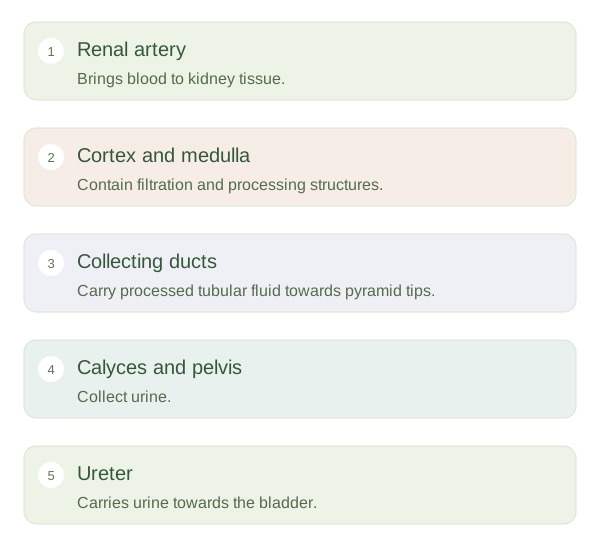
Kidneys process blood within tissue and collect urine into a separate drainage route.
 Open diagram larger ↗
Blood supply supports processing
A renal artery brings blood from the aorta to each kidney. Branches progressively divide until blood reaches afferent arterioles supplying glomeruli, the capillary tufts where filtration begins. Blood leaves each glomerulus through an efferent arteriole and then passes into another capillary network around tubules. This arrangement supports both filtration and later exchange. Renal veins return blood towards the inferior vena cava. The kidneys receive substantial blood flow because they continuously regulate the composition of the internal environment.
Urine takes another route
Fluid remaining after tubular processing enters collecting ducts, then drains from pyramid tips into minor calyces. These join larger spaces and the , which leads into the ureter. The renal pelvis is a collecting funnel, not a blood-filtering cavity. Blood and urine therefore follow neighbouring but distinct pathways. A blockage in the urine drainage route can raise pressure upstream and disturb kidney function even though the renal artery is still present. Likewise, reduced blood supply can impair function without an initial ureter blockage. Learning both routes explains why the kidney is not simply a sieve connected to a waste pipe: it contains a precisely organised circulation, processing tubules and a collecting system.