A microscopic functional unit
A consists of a renal corpuscle and a connected tubule. The corpuscle contains a surrounded by Bowman’s capsule. Pressure drives water and small dissolved substances from glomerular blood into the capsule’s space, creating . Blood cells and most large proteins normally remain in the circulation. Filtrate is not finished urine: it contains useful substances that the body must reclaim, as well as material that may ultimately be excreted.
A nephron filters first, then selectively modifies the resulting fluid.
 Open diagram larger ↗
A sequence of specialised segments
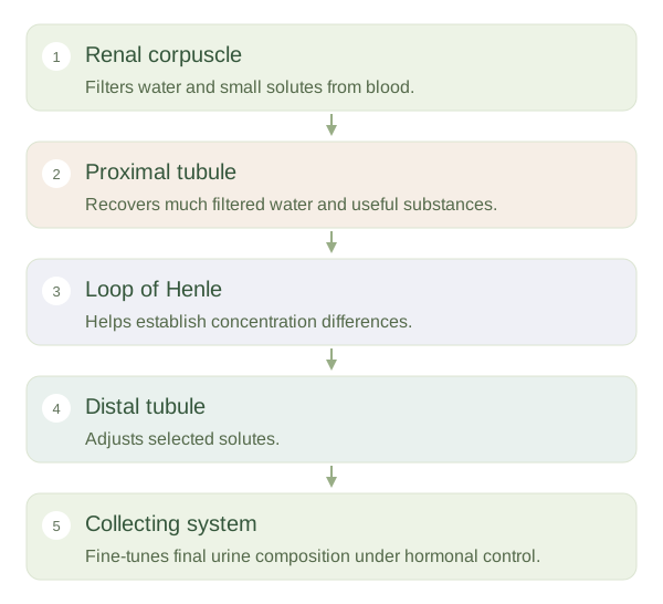
Fluid enters the proximal tubule, where much water and many useful solutes are reabsorbed. It then passes through the , with descending and ascending limbs that have different permeability and transport properties. These differences help establish the kidney’s internal concentration gradient. The distal tubule and collecting system make further adjustments. Different segments express different transport proteins, so the cannot be understood as one uniform pipe with identical behaviour throughout.
Tubules and vessels work together
After filtration, blood enters capillaries around the tubules. These receive reabsorbed water and solutes and supply substances that may be secreted into tubular fluid. Long loops and associated vessels help maintain concentration differences in the medulla. Collecting ducts receive fluid from multiple nephrons and respond to hormones that influence final water and electrolyte handling. Strictly, the collecting duct system is anatomically distinct from an individual , although it is essential to urine formation. The useful overall map is filtration at the corpuscle, bulk recovery early in the tubule, gradient-building in the loop and regulated adjustment downstream. This arrangement lets the kidneys discard wastes while retaining most of the water and valuable molecules initially filtered.