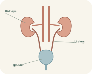
Two delivery tubes, one storage organ
Each kidney drains into a , a muscular tube leading towards the bladder. The ureters transport urine using coordinated smooth-muscle contractions, with pressure and gravity also contributing. They do not rely only on a person standing upright. As a ureter enters the bladder wall, its oblique course helps limit backward flow when bladder pressure rises. This is a functional anti-reflux arrangement, not a separate mechanical valve that opens and closes like a tap.
Ureters deliver urine to the bladder; the urethra carries it outside.
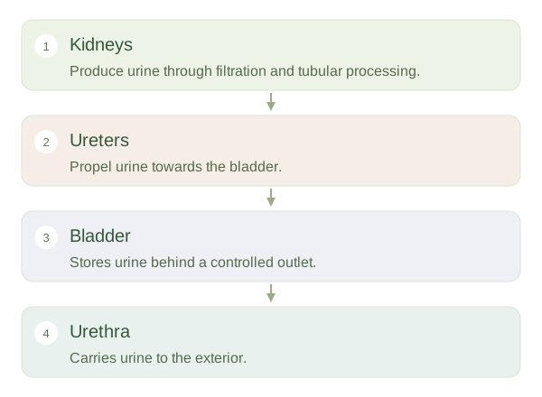 Open diagram larger ↗
Storage requires a specialised wall
The bladder sits in the pelvis and expands as it fills. Its inner urothelium, also called transitional epithelium, can accommodate stretching while helping maintain a barrier against urine. The muscular wall contains the . During filling, coordinated neural activity allows the bladder to hold increasing volume without a matching large rise in pressure. Pelvic support and outlet control also matter. The bladder stores urine already produced by the kidneys; it is not the main site where blood is filtered or urine concentration is adjusted.
One outlet with variable anatomy
The carries urine from the bladder to the outside. Its length and relationship to reproductive structures differ between anatomical patterns. A shorter urethra gives organisms a shorter potential route towards the bladder, contributing to differences in infection risk without being the only factor. The longer urethra associated with a penis also carries semen at different times. Ureters and urethra are easily confused because their names are similar, but their jobs are distinct. Following the route in order prevents the confusion: kidneys produce and modify urine, ureters deliver it, the bladder stores it and the urethra provides the final exit.