Filling is an actively coordinated state
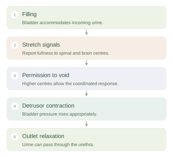
As urine enters the bladder, its wall accommodates increasing volume. During storage, activity remains suitably restrained while the outlet and pelvic-floor support help maintain . Stretch-sensitive signals report filling through nerves to spinal and higher centres. Awareness of bladder fullness develops gradually rather than appearing only when the organ is completely full. A healthy system can usually delay emptying until an appropriate setting, but the capacity for delay varies with age and circumstances.
Urination requires bladder contraction and outlet relaxation to work together.
 Open diagram larger ↗
Emptying needs the opposite pattern
When voiding is permitted, brainstem and spinal circuits coordinate contraction with relaxation of the outlet. Parasympathetic pathways are important in activating the bladder muscle. The is skeletal muscle under somatic neural control, providing an important voluntary influence. Smooth muscle at the bladder outlet also contributes, with anatomical differences between people. Effective emptying requires coordination rather than simply making the bladder squeeze as hard as possible against a closed exit.
Control spans several levels
Signals travel between the bladder, sacral spinal cord, brainstem and cerebral networks. Damage or disruption at different levels can produce different problems, including urgency, leakage, retention or poorly coordinated emptying. Pelvic-floor weakness can also affect without being the same as a failure of bladder sensation. Urine production is a separate upstream process: making more urine increases filling, but does not by itself explain every control problem. A useful analogy is a reservoir with both a pump and a gate. Safe emptying needs the pump to act when the gate is appropriately open, while safe storage needs adequate capacity and outlet support. Understanding this coordinated switch connects anatomy, autonomic regulation and conscious behaviour without reducing urination to one isolated reflex.