A small link beneath the brain
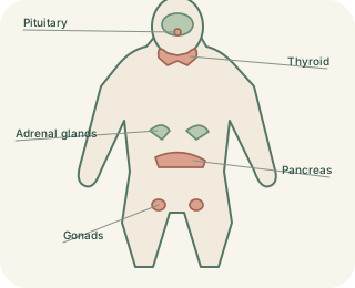
The is a brain region involved in temperature, hunger, water balance and many other survival functions. Just below it lies the pituitary gland, connected by a stalk. Together they translate information about the body into hormonal instructions. Calling the pituitary a master gland can help initially, but it is not an independent commander: the hypothalamus, target glands and feedback signals all influence its activity.
Two routes through the pituitary
 Open diagram larger ↗
The anterior pathway
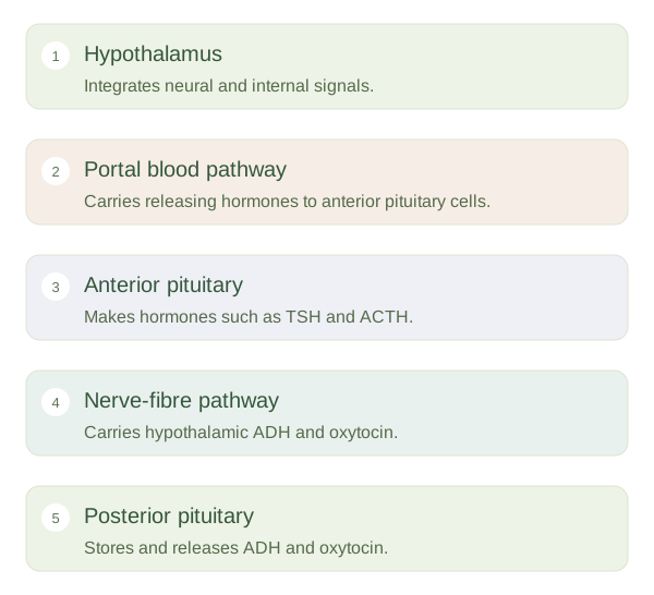
The contains endocrine cells that manufacture their own hormones. The controls these cells through releasing and inhibiting hormones delivered in a specialised portal blood supply. An example is TRH stimulating TSH release, which then stimulates the thyroid. Other anterior pituitary hormones include ACTH, which influences the adrenal cortex; FSH and LH, which regulate gonadal functions; growth hormone; and prolactin. Different cell populations make these different messages.
The posterior pathway
The is organised differently. Neurons in the make antidiuretic hormone, also called or vasopressin, and oxytocin. These hormones travel down nerve fibres and are stored and released from terminals in the posterior pituitary. ADH helps the kidneys conserve water when appropriate. Oxytocin contributes to uterine contractions and milk ejection. The posterior pituitary releases these hormones, but the hypothalamic neurons are their source.
Coordination rather than isolation
This arrangement allows nervous and endocrine control to cooperate. For instance, changes in blood concentration can be detected centrally and lead to altered release. Suckling can trigger neural signals that promote oxytocin release. Hormones from downstream glands also feed information back to the brain and pituitary. Understanding the two pathways prevents a common confusion: not every hormone released by the pituitary was made by pituitary endocrine cells.