Calcium has two lives
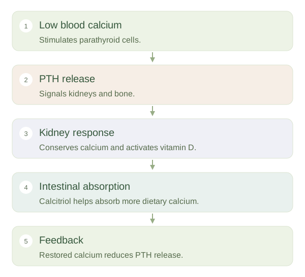
Most body calcium is stored in the skeleton, helping make bone strong. A much smaller amount circulates in blood and tissue fluid, where calcium ions support nerve signalling, muscle contraction and blood clotting. These jobs require careful regulation of the free, concentration. The small parathyroid glands, usually found on the back of the thyroid, detect changes in blood calcium and release parathyroid hormone, or , when it falls.
Responding to low calcium
 Open diagram larger ↗
Kidneys and vitamin D
tells the kidneys to conserve more calcium rather than lose it in urine. It also promotes phosphate excretion and encourages production of , the active form of vitamin D. Calcitriol increases the intestine's ability to absorb dietary calcium. These are connected responses: a message from the neck changes kidney handling, which then changes intestinal absorption. PTH therefore affects the gut mainly through the active vitamin D pathway rather than acting as a digestive enzyme.
Drawing on the bone store
Bone is continually remodelled by cells that remove old material and cells that build new material. can increase calcium release from bone through signals involving bone-forming cells and bone-resorbing cells. This helps defend the blood calcium level, but prolonged excessive PTH can weaken the skeleton. A normal concentration in blood does not necessarily prove that the body has plentiful calcium stores, because regulation may maintain blood levels partly at the expense of bone.
Closing the loop
As blood calcium rises towards a suitable range, parathyroid cells reduce release. This is negative feedback. The thyroid and parathyroids are close anatomically but have different main functions: thyroid hormones influence metabolism, whereas PTH is central to calcium regulation. Calcitonin can lower calcium in some circumstances, but it has a less dominant role in everyday adult calcium control than PTH and vitamin D. Several organs cooperate to keep this small circulating calcium pool usable.