Production in the testes
The testes lie within the scrotum, outside the main abdominal cavity. This position helps maintain conditions slightly cooler than core body temperature, supporting normal sperm production. Within each testis, tightly coiled seminiferous tubules contain developing sperm cells and supporting Sertoli cells. Leydig cells between the tubules produce testosterone. Sperm production and hormone production therefore occur in neighbouring but different cell populations.
The sperm transport route
 Open diagram larger ↗
Maturation and the duct route
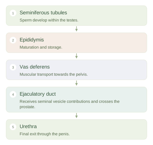
Newly formed sperm move into the , a long coiled duct along the back of the testis. There they mature and gain functions needed later in reproduction; the lower part also stores them. During ejaculation, smooth muscle contractions propel sperm along the ductus deferens, also called the . It travels within the spermatic cord and enters the pelvis. Its connection with a seminal vesicle duct forms an ejaculatory duct that passes through the prostate into the urethra.
Adding the fluid
is more than sperm. Seminal vesicles and the prostate add much of its fluid, including substances that support sperm function and transport. Bulbourethral glands contribute mucus before ejaculation. The urethra passes through the penis and provides the final exit route. It also carries urine at other times, but coordinated muscular and neural activity normally prevents urine and semen being expelled together. The ducts themselves move sperm; sperm do not swim the whole route from the testis unaided.
Structure explains function
Blocking the prevents sperm reaching the ejaculate without directly stopping testosterone production, because Leydig cells remain supplied by blood. This illustrates why understanding the pathway is more useful than memorising names alone. Reproductive anatomy varies between individuals and after surgery. The structures described here are a common anatomical arrangement; their presence does not determine a person's gender identity, fertility or individual sexual experience.