A layered organ
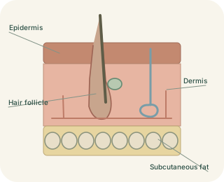
Skin is an organ with two main layers: the outside and the underneath. Beneath them lies the , or subcutaneous tissue, which connects skin to deeper structures and commonly contains fat. This lower tissue supports insulation, cushioning and energy storage, but it is usually distinguished from the skin itself. Different body regions have different thicknesses and specialised structures, so one section cannot represent every patch of skin.
Skin from surface to depth
 Open diagram larger ↗
The renewing epidermis
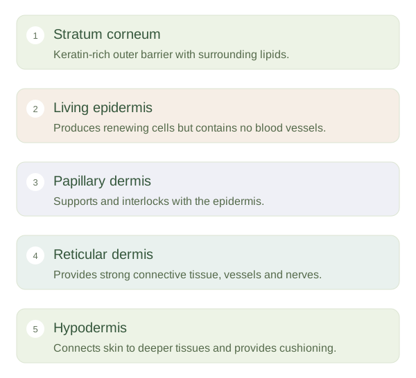
The is a stratified squamous epithelium, meaning it contains multiple layers of cells that become flattened towards the surface. Most are keratinocytes. New cells arise near the base, move outward and gradually form a tough, keratin-rich surface. The outer stratum corneum contains flattened cells and surrounding lipids that help limit water loss and entry of harmful substances. The epidermis has no blood vessels; nutrients diffuse from vessels in the underlying . Melanocytes produce pigment, while other cells contribute to immune surveillance and sensation.
The supporting dermis
The contains connective tissue rich in collagen and elastic fibres, giving skin tensile strength and flexibility. Its upper papillary region interlocks with the , while its deeper reticular region contains denser connective tissue. Blood vessels support nutrition and temperature regulation. Nerves and specialised receptors detect touch and other stimuli, and many hair follicles and glands extend into this layer. A cut reaching the dermis may therefore bleed, unlike a superficial injury confined to avascular epidermis.
Structure fits the location
Palms and soles have a particularly thick and an additional clear layer, the stratum lucidum, but lack hair follicles. Most other skin is classified as thin skin even when the underlying is substantial. The labels refer principally to epidermal structure, not overall body thickness. Understanding these layers helps explain why depth matters in injuries: damage can affect not only the barrier but also the structures that nourish, sense and regenerate the surface.