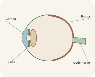
A living optical system
The eye gathers light and directs it towards the , the light-sensitive tissue lining its back. The transparent forms the front window and supplies much of the eye’s light-bending power. Light then passes through the , the opening in the coloured iris. The iris changes pupil size to help regulate incoming light. Behind it sits the clear lens. Aqueous humour fills the anterior spaces, while a gel called vitreous humour occupies much of the larger space behind the lens.
The route of light
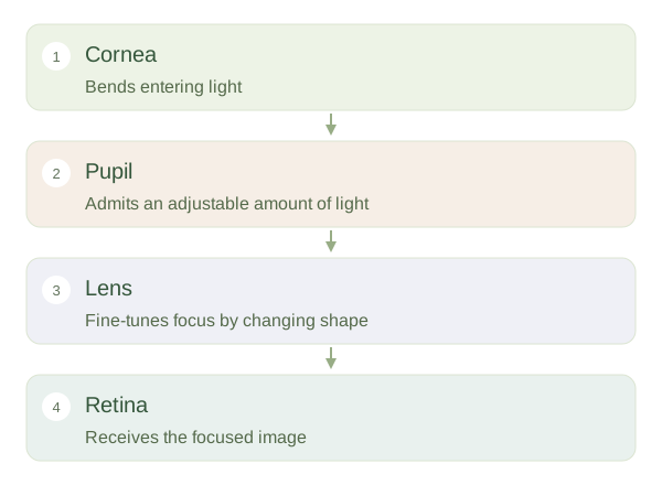 Open diagram larger ↗
Changing the focus
Light from a nearby object spreads out more before reaching the eye than light from a distant object. To focus a nearby object on the , the lens becomes rounder and bends light more strongly. The ciliary muscle contracts, reducing tension in the supporting zonular fibres so the elastic lens can change shape. For distant viewing, these fibres pull the lens flatter. This adjustment is . It changes the optical focus; the retina does not move forwards and backwards like a camera screen.
Why clear focus matters
A sharp retinal image allows neighbouring points in a scene to stimulate distinct groups of receptors. In short-sightedness, distant images focus in front of the when is relaxed. In long-sightedness, the relaxed optical system would focus behind it. Corrective lenses alter incoming light before it enters the eye. With age, the natural lens becomes less able to change shape, making near focus harder; this is presbyopia. Focusing and sensing are different jobs: transparent tissues focus light, while retinal cells convert it into signals. Both functions are necessary for useful vision.