Where light becomes information
The retina is a thin part of the nervous system lining the inside of the eye. Its photoreceptors respond to light using specialised molecules that change when they absorb photons. Rods are highly sensitive and help us see in dim conditions, although they do not provide normal colour discrimination. Cones support colour vision and fine detail in brighter light. Humans usually have three classes with overlapping wavelength sensitivities. Colour comes from comparing their activity, rather than assigning each cone a single painted colour.
Retinal signal processing
 Open diagram larger ↗
A network before the brain
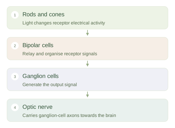
Photoreceptors communicate through bipolar cells to ganglion cells, with other retinal neurons modifying these connections. This network emphasises useful differences, such as contrast and changing edges. Ganglion-cell axons gather to form the optic nerve. Light causes photoreceptors to become more electrically negative and changes their transmitter release; the retina then converts these changes into patterns of ganglion-cell activity. The eye therefore sends processed information, not a tiny photographic file. Signals from many receptors may converge, trading fine detail for sensitivity.
Special places on the retina
The is a small central retinal region with densely packed cones and connections suited to detailed vision. You aim an object towards the fovea when reading a word or inspecting a face. Peripheral retina is better suited to detecting dim light and movement over a wider area. At the optic disc, axons leave the eye and there are no photoreceptors, creating a normal blind spot. Nearby visual information and input from the other eye usually prevent awareness of that gap. This organisation explains why looking directly at a very faint star can make it harder to see.