One scene, two retinal images
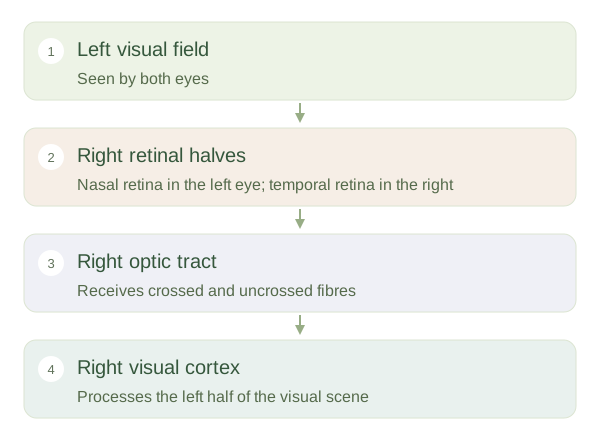
Each eye views much of the same scene from a slightly different position. The cornea and lens form an inverted image on each retina, but perception is not an upside-down picture that needs physical rotation. Neural circuits interpret the arrangement of activity. Light from the left half of the scene reaches the right halves of both retinas. Consequently, visual pathways are organised around halves of the visual world, rather than simply sending the whole left eye to the right brain.
Left visual field to right visual cortex
 Open diagram larger ↗
The partial crossing
Retinal ganglion-cell axons form the optic nerves. At the , axons from the nasal half of each retina cross to the opposite side; those from the temporal half remain on their original side. Each optic tract therefore carries information about the opposite from both eyes. Most fibres relay in the thalamus before travelling through optic radiations to the primary visual cortex in the occipital lobe. Other fibres help control pupil responses and eye movements without requiring conscious visual awareness.
Constructing depth
The brain compares small differences between the two retinal images, called , to estimate relative distance. This is . Eye alignment is important because corresponding features must be matched. However, depth does not depend on two eyes alone. Overlap, perspective, relative size, shading and the movement of objects across the scene also supply useful clues. You can still judge many distances with one eye closed, although close, precise tasks may become harder. Vision is therefore an active interpretation combining retinal signals with context, learned expectations and information about where the eyes are pointing.