A covering with several layers
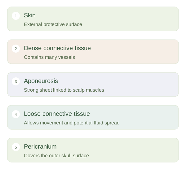
The scalp covers the upper skull and consists of skin, dense connective tissue, a broad , loose connective tissue and the . The aponeurosis is a tough tendon-like sheet linking parts of the occipitofrontalis muscle. Skin, dense tissue and the aponeurosis move together over the looser layer beneath. The pericranium is the outer connective-tissue covering of the skull bones. These layers help explain how the scalp can slide slightly while remaining securely attached around its margins and along particular underlying structures.
Scalp from surface to skull
 Open diagram larger ↗
Why scalp wounds can bleed
Many scalp vessels run in the dense connective-tissue layer. This tissue can hold cut vessels open, contributing to conspicuous bleeding after a scalp wound. The amount of visible blood alone does not reliably indicate how deep the underlying injury is. Loose connective tissue beneath the allows fluid or blood to spread across a broader area. It also contains communicating veins, which provide potential connections between superficial tissues and intracranial venous channels. The anatomy explains possible routes of spread without meaning that every minor scalp problem reaches the brain.
The face has a different arrangement
Facial tissues also include skin, fat, vessels and muscles, but many expression muscles attach into skin or nearby soft tissue rather than ending only on another bone. This lets contraction move the surface into a smile or a frown. Facial sensory supply mainly comes from branches of the , while the powers most expression muscles. Distinguishing sensation from movement prevents a common confusion: a nerve called facial is not responsible for all facial feeling. Layers, attachments and different nerve functions together explain how the head covering protects, moves and communicates.