A protected space, not an empty socket
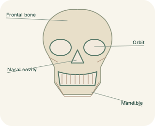
The is a bony cavity containing the eyeball, extraocular muscles, nerves, vessels, fat and much of the tear-producing apparatus. Its rim helps protect the eye, while fat cushions structures and allows movement. Several skull and facial bones contribute to the orbital walls. The medial wall and floor are relatively thin and lie beside the ethmoidal air cells and maxillary sinus respectively. These close neighbours explain why disease or injury can involve both an orbit and an adjacent sinus without being confined to the visible eye.
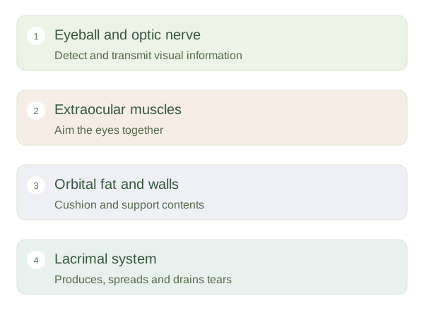
Orbit: functions of its contents
 Open diagram larger ↗
Moving the eye precisely
Six extraocular muscles rotate the eyeball. Four rectus muscles and two oblique muscles work in coordinated pairs so both eyes aim at the same target. Most receive motor supply from the oculomotor nerve; the superior oblique uses the trochlear nerve and the lateral rectus uses the abducens nerve. The optic nerve carries visual information rather than moving the eye. An eye can therefore have a movement problem even when the retina still detects light. Correct alignment supports a single image and accurate comparison between the two eyes.
Tears and routes through bone
The lies towards the upper outer and releases the watery component of tears. Blinking spreads tears across the front of the eye, after which fluid drains towards openings near the inner eyelid margins and down the into the nose. This helps explain a runny nose during crying. At the back of the orbit, openings including the optic canal and superior orbital fissure provide nerve and vessel routes. Thinking in terms of contents, neighbouring spaces and passageways makes the orbit easier to understand than treating it as a single solid structure.