Two joints acting together
The mandible meets the temporal bone at a on each side of the head. Because the two joints belong to the same lower jaw, their movements are linked. A fibrous separates joint spaces and helps accommodate movement between the mandibular and temporal surface. The covering tissue differs from the typical hyaline cartilage found in many other synovial joints. Capsules and ligaments support the joints, while muscles actively guide the position of the jaw during eating and speaking.
Jaw opening and control
 Open diagram larger ↗
Opening is more than a hinge
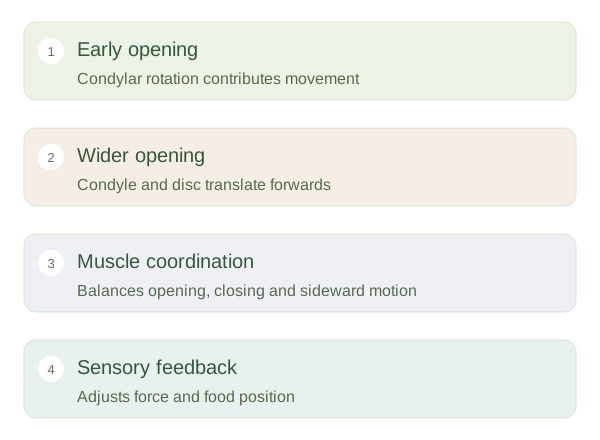
Early mouth opening includes rotation of the mandibular . Wider opening also involves forward of the condyle and disc along the temporal articular surface. Side-to-side chewing combines movements at both joints. Masseter, temporalis and the medial pterygoid are important jaw closers. The lateral pterygoid helps guide forward and sideward movements, while muscles associated with the hyoid can assist opening when their other attachments are stabilised. The main chewing muscles receive motor supply through the mandibular division of the trigeminal nerve, rather than the facial nerve.
A coordinated chewing system
Teeth break food into smaller pieces, but they cannot perform this task efficiently without controlled jaw movement. Sensory information from muscles, joints and tissues around the teeth helps regulate bite force and position. The tongue and cheeks continually reposition food, so chewing is a coordinated activity involving more than the joint itself. A painless click does not by itself establish a serious disorder, and jaw symptoms can arise from muscles as well as the joint. For learning anatomy, the important distinction is between the joint’s available movement and the muscular control that uses it.