A busy connection between head and chest
The neck carries air, food, blood and nerve signals between the head and trunk while supporting a mobile head. Its contents are not arranged as loose items in one open space. Connective-tissue sheets called wrap muscles, vessels and organs into related compartments. Superficial tissues contain skin, fat and the thin platysma muscle. Deeper fascial layers surround major muscle groups and the visceral structures near the centre. Their boundaries provide a useful organising map, although real anatomy has variations and communicating routes.
Organising the neck
 Open diagram larger ↗
Compartments and their contents
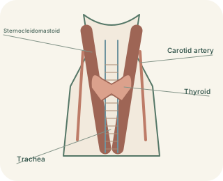
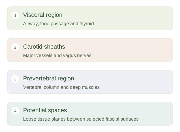
The central visceral region contains structures such as the pharynx, larynx, trachea, oesophagus and thyroid. Behind it are the vertebral column and deep muscles, associated with the . On either side, a encloses major vessels and the vagus nerve. The sternocleidomastoid muscles provide visible landmarks that divide each side into anterior and posterior triangles. These triangles are surface descriptions, not sealed boxes. They help locate structures relative to muscles, the midline and the clavicle without replacing the deeper compartment model.
Why potential spaces matter
Some fascial surfaces lie close together with loose tissue between them, creating potential spaces. Fluid, blood or infection may separate these layers and travel beyond the original site. A retropharyngeal collection, for example, lies behind the pharynx and may affect swallowing or nearby airways. Certain deeper pathways can communicate towards the chest. This explains why the position of swelling can matter as much as its external size. It does not mean all neck discomfort represents deep disease. The anatomical lesson is that fascial boundaries guide both normal organisation and possible routes of spread.