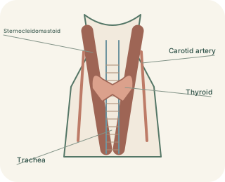
A supported entrance to the airway
The connects the pharynx above with the trachea below. Cartilages provide a framework that can change shape without collapsing during normal breathing. The thyroid cartilage forms much of the front and sides, while the ring-shaped cricoid lies below it. Arytenoid cartilages towards the back help position the vocal folds. The epiglottis lies above the entrance and contributes to protection during swallowing. These structures form a mobile valve system, not merely a box that happens to make sound.
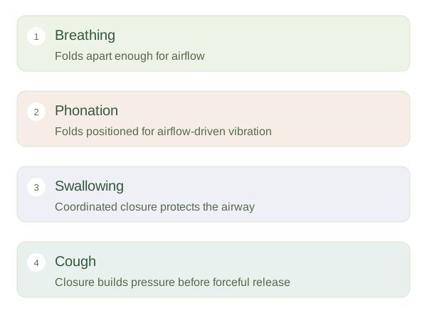
Laryngeal settings for different tasks
 Open diagram larger ↗
How voice is generated
The true vocal folds extend within the , with a gap called the between them. For , muscles bring the folds into an appropriate position and air from the lungs drives repeated vibration. Changing fold tension, length and the pattern of closure influences pitch and voice quality. The mouth, tongue, lips and resonating spaces then shape this sound into speech. The vocal folds do not repeatedly contract once for each sound-wave cycle; airflow and tissue mechanics sustain the rapid vibration after muscles set the conditions.
One structure, several coordinated jobs
During quiet breathing, the vocal folds are sufficiently apart to allow airflow. During swallowing, laryngeal closure helps protect the lower airway. Closure can also build pressure for a cough before the folds open and air is expelled. Most intrinsic laryngeal muscles receive motor supply from recurrent laryngeal branches of the vagus nerve; cricothyroid receives supply through the external superior laryngeal branch. Sensory pathways are also essential for protective reflexes. Voice changes therefore may reflect altered tissue vibration, impaired muscle control or other causes. Anatomy explains the possibilities without identifying a diagnosis from sound alone.