A map on the body surface
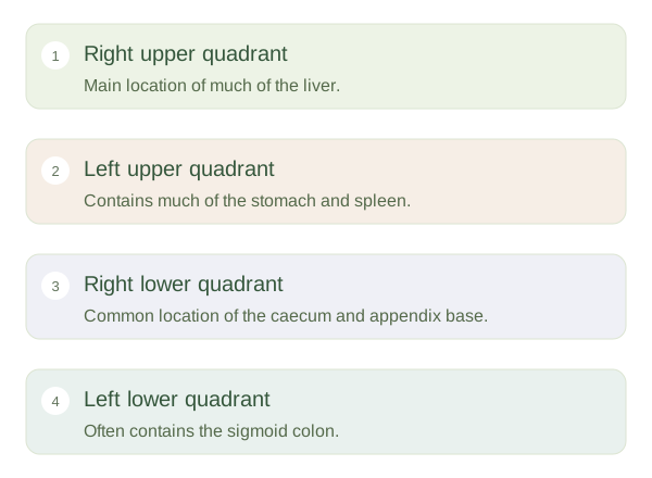
The abdomen lies between the diaphragm above and the pelvis below. Its front wall gives us a useful surface map of deeper organs. One simple scheme divides it into four quadrants using a vertical midline and a horizontal line through the . These are the right upper, left upper, right lower and left lower quadrants. Right and left always refer to the person being described, not the viewer. This matters when looking at another person or an anatomical illustration: the person’s right appears on your left in a front view.
Abdominal Regions and Surface Landmarks — concept map
 Open diagram larger ↗
A more detailed grid
A nine-region scheme uses two vertical and two horizontal reference lines. The upper row contains right hypochondriac, epigastric and left hypochondriac regions. The middle row contains right lumbar, umbilical and left lumbar regions. The lower row contains right iliac, hypogastric and left iliac regions. You do not need to memorise the names immediately to understand their purpose: they let people describe a more specific area. The rib margins, sternum, hip bones and pubic region provide additional landmarks. These external points are guides rather than windows into the abdominal cavity.
Organs do not fit neatly in boxes
The liver lies mainly in the right upper abdomen, while much of the stomach is on the left. Bowel loops occupy several regions, and their positions change with movement and filling. Breathing moves the diaphragm and some underlying organs. Body shape, pregnancy, previous surgery and normal variation can also alter relationships. Pain may be referred away from its source, so an area of discomfort does not identify an organ with certainty. Surface maps are most useful when combined with knowledge of organ depth and connections. Their role is to communicate location clearly, not to turn one symptom location into a diagnosis.