A flexible protective wall
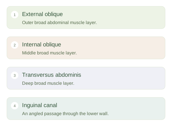
The abdominal wall contains skin, connective tissue, muscle and deeper lining tissues. Unlike the chest, much of it has no rigid bony cage. Three broad paired muscles form its sides: external oblique, internal oblique and transversus abdominis. Their fibres run in different directions, making a strong flexible arrangement. Rectus abdominis runs vertically near the midline within a connective-tissue sheath. The muscles help bend or rotate the trunk, resist movement and compress the abdominal contents. Their broad tendinous sheets meet at the , the connective-tissue seam in the middle.
Abdominal Wall and Inguinal Canal — concept map
 Open diagram larger ↗
A passage through the layers
The is an oblique passage in the lower abdominal wall, just above the medial part of the inguinal ligament. It extends between a deep ring and a superficial ring. It carries the spermatic cord in typical male anatomy and the round ligament of the uterus in typical female anatomy. The ilioinguinal nerve also passes through part of the canal but does not enter through its deep ring. An angled route through several layers helps preserve wall strength, although any necessary opening creates a potential weak region.
Why hernias occur here
A is a protrusion of tissue through a weakness or opening in the wall containing it. An indirect inguinal hernia enters through the deep ring, lateral to the inferior epigastric vessels. A direct inguinal hernia pushes through a weakened posterior wall medial to those vessels. Both differ from a femoral hernia, which passes below the inguinal ligament through another route. These names describe anatomy, not severity. Increased abdominal pressure and tissue properties influence how weaknesses become apparent. The essential idea is that a wall must balance strength with the need for structures to pass through it; the canal illustrates that compromise.