One membrane, two descriptions
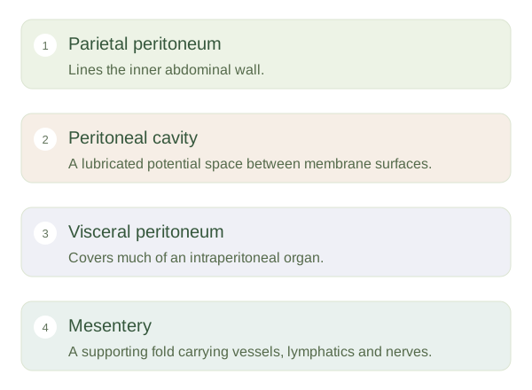
The is a thin serous membrane associated with the abdominal cavity. Parietal peritoneum lines the inner wall, while visceral peritoneum covers the surfaces of many organs. These are continuous parts of a single membrane, not unrelated bags. Between opposing surfaces is a potential space, the peritoneal cavity, containing a small amount of lubricating fluid. This lets organs slide during breathing, digestion and movement. The cavity is not the inside of the bowel, and abdominal organs do not normally float freely inside a large empty fluid-filled chamber.
Peritoneum and Mesenteries — concept map
 Open diagram larger ↗
Folds carry essential connections
A is a double layer of connecting an organ to a supporting wall or another structure. Within it travel arteries, veins, lymphatics and nerves. The mesentery of the small intestine suspends much of the jejunum and ileum from the posterior abdominal wall. It allows movement without disconnecting their supply. Omenta are specialised peritoneal folds associated with the stomach. The greater omentum hangs over bowel loops and contains fat and immune-related tissues. These folds are therefore active routes and supporting structures, rather than disposable packaging around the digestive tract.
Different organ positions
Intraperitoneal organs are largely covered by visceral and often have a . organs lie behind the parietal peritoneum on the posterior wall. The kidneys are retroperitoneal; most of the pancreas and duodenum are also behind the peritoneal lining in adults. An organ can be part of the digestive system without being intraperitoneal. The distinction helps explain mobility, surgical access and how fluid or inflammation can spread. Parietal irritation is often more precisely localised than pain from visceral tissues, which is usually harder to pinpoint. Understanding the membrane’s reflections makes an apparently crowded abdomen much easier to organise.