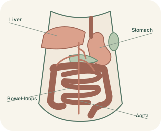
Start beneath the diaphragm
The upper abdomen sits directly below the diaphragm. The liver occupies much of the right upper region and extends towards the left. The stomach lies mainly on the left, with its upper part beneath the left diaphragm. The lies farther to the left and posterolateral to the stomach, protected partly by lower ribs. These relationships describe the person’s own right and left. Breathing shifts the diaphragm and can move the liver and spleen slightly. Organs also vary in shape and size, so a diagram should be treated as an orientation map rather than a perfectly fixed arrangement.
Upper Abdominal Organ Relationships — concept map
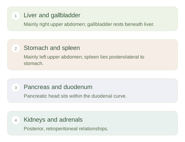 Open diagram larger ↗
Look behind and below
The pancreas lies largely behind the stomach. Its head fits into the curve of the , while its tail extends towards the . The pancreatic duct carries digestive secretions towards the duodenum. The gallbladder rests against the underside of the liver and stores bile made by the liver. Its drainage joins the biliary route towards the duodenum. The kidneys lie farther posteriorly, behind the peritoneum; the right kidney is commonly slightly lower than the left. The adrenal glands sit near their upper poles and have endocrine, not urine-forming, functions.
Connections make the map useful
Spatial proximity does not mean identical function. The filters blood and supports immunity, whereas the stomach processes swallowed food. The liver handles absorbed substances and produces bile, while the pancreas contributes digestive enzymes and hormones. Their ducts, vessels and connective-tissue attachments create pathways between neighbouring structures. This helps explain why a problem in one region can affect more than one organ, although anatomy alone cannot diagnose a symptom. On cross-sectional images, the stomach, pancreas, liver, kidneys and major vessels are useful landmarks for orienting the viewer. First identify front versus back and the person’s right versus left, then trace relationships.