Arteries follow major territories
The abdominal aorta runs in front of the vertebral column, slightly left of the midline. Three major unpaired branches supply much of the digestive tract: the coeliac trunk, superior mesenteric artery and inferior mesenteric artery. Their territories broadly follow the foregut, midgut and hindgut divisions established during development. The coeliac trunk supplies structures including the stomach, liver and spleen. The superior mesenteric artery supplies much of the small intestine and proximal large intestine, while the inferior mesenteric artery supplies more distal large bowel. Overlapping connections can link neighbouring arterial territories.
Abdominal Arteries, Portal Vein and Venous Drainage — concept map
 Open diagram larger ↗
A second capillary bed
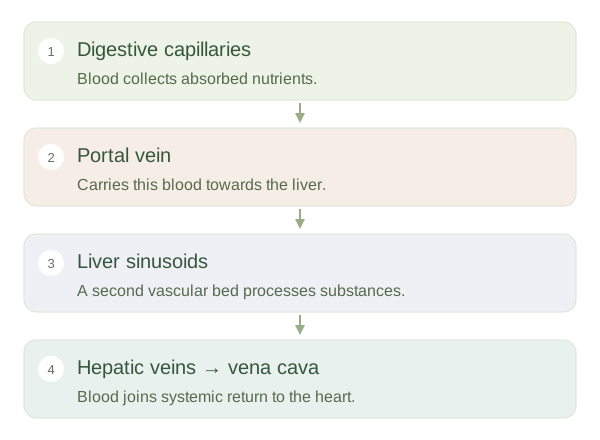
Venous blood from much of the digestive tract, spleen and pancreas enters the hepatic portal system. The portal vein is usually formed by the union of the superior mesenteric and splenic veins; the inferior mesenteric vein commonly drains into the splenic vein. Portal blood reaches liver sinusoids, specialised small vascular channels, before leaving through hepatic veins to the . This lets the liver process absorbed nutrients and other substances before they reach the wider circulation. The liver also receives oxygenated blood from the hepatic artery, so portal blood is only part of its dual supply.
Not every abdominal vein is portal
The ascends on the right side of the aorta and carries systemic venous blood towards the heart. The renal veins drain the kidneys into this systemic route rather than through the portal vein. Portal and systemic veins have connections at some sites. If portal pressure rises, these alternative pathways can enlarge, but they do not make liver passage unnecessary under normal conditions. The important distinction is between route and oxygen content: a portal vein is named for connecting two capillary networks, not for carrying a special kind of oxygen-free blood.