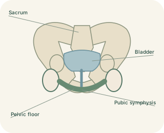
A ring built from several bones
The bony pelvis consists of the two hip bones, the sacrum and the coccyx. Each adult hip bone forms from the fusion of three parts: ilium, ischium and pubis. They meet around the , the socket receiving the femoral head. The broad ilium forms the upper wing, the ischium contributes to the lower posterior region, and the pubis lies towards the front. The ischial tuberosities bear much of the body’s weight when sitting. These components create a continuous ring rather than two independent supports beneath the trunk.
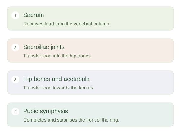
Pelvic Bones and Joints — concept map
 Open diagram larger ↗
Joints distribute forces
Behind, the sacroiliac joints connect the sacrum with the ilia. In front, the joins the pubic bones through fibrocartilage. Strong ligaments reinforce the ring and limit movement. Weight from the spine enters through the sacrum and passes towards the hip joints, then into the femurs during standing. When sitting, forces are redirected towards the ischial tuberosities. Small movements at pelvic joints are normal, but this region prioritises stability more than the freely moving shoulder girdle. The hip joint is distinct from the : one joins pelvis to femur, the other joins pelvis to sacrum.
Cavity and variation
The pelvic brim separates the greater pelvis above from the lesser, or true, pelvis below. The lesser pelvis contains structures such as the bladder and rectum, along with reproductive organs appropriate to the person’s anatomy. The shape and dimensions of the pelvic opening vary between individuals. Typical sex-related patterns exist, but they overlap and do not determine every individual feature. During childbirth, the dimensions of the bony passage interact with fetal size, position and soft tissues. The pelvis therefore combines three roles: mechanical load transfer, protection of organs and formation of a passage through the lower trunk.