Begin with front and back
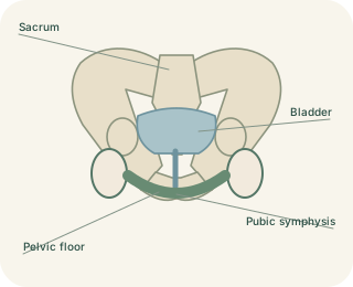
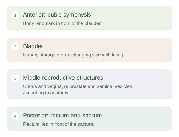
The urinary bladder lies towards the front of the true pelvis, behind the . The rectum lies farther back, in front of the sacrum and coccyx. Between and around them lie reproductive structures whose arrangement depends on anatomy. These relationships are easiest to picture in a side view. A full bladder expands upwards and may extend into the lower abdomen, so pelvic organs do not remain the same size or position throughout the day. The rectum also changes shape as it fills, while supportive tissues help maintain the overall arrangement.
Bladder, Rectum and Reproductive Organ Relationships — concept map
 Open diagram larger ↗
Two common anatomical arrangements
Where a uterus and vagina are present, the uterus commonly lies above the vagina and between bladder and rectum. It often tilts forwards, but normal positions vary. The vagina passes behind the bladder and urethra and in front of the rectum. Where a is present, it lies below the bladder, surrounding the proximal urethra, and in front of the rectum. Seminal vesicles lie behind the bladder. The testes lie outside the pelvic cavity in the scrotum, although their ducts and vessels connect them with structures higher in the body.
Shared space, distinct pathways
Urine leaves through the urethra; stool passes through the rectum and anal canal. These remain separate routes even where organs lie close together. Peritoneal reflections create recesses between some pelvic organs. In typical uterine anatomy, the lies between uterus and rectum; in typical anatomy, a rectovesical pouch lies between bladder and rectum. Such relationships influence where fluid may collect. Organ enlargement, pregnancy or support changes can alter the space available to neighbours. This explains possible interactions without implying that every urinary or bowel symptom arises from a nearby reproductive organ. Anatomical proximity is a clue to relationships, not a diagnosis.